- 1. Features of Gas Exchange Surfaces
- 2. Gases in Atmospheric Air
- 3. Differences Between Inspired and Expired Air
- 4. The Gas Exchange System — Structures to Identify
- 5. The Alveoli — The Gas Exchange Surface
- 6. The Breathing Mechanism
- 7. Effect of Physical Activity on Breathing
- 8. Protecting the Gas Exchange System
IGCSE Biology | Life Processes
Every cell in your body needs oxygen to release energy from food, and it produces carbon dioxide as a waste product. Your gas exchange system brings oxygen from the air into your blood and removes carbon dioxide from your blood back into the air. This topic covers how that system is structured, how it works, and how your body protects it.
1. Features of Gas Exchange Surfaces #
For gases to move between the air and the blood quickly enough to supply the whole body, the gas exchange surface must have three key features:
| Feature | Why it is needed |
|---|---|
| Large surface area | More surface means more gas can cross at the same time. This makes gas exchange fast enough to meet the body’s needs. |
| Thin surface | Gases diffuse across a short distance very quickly. A thicker surface would slow this down too much. |
| Good blood supply and air supply | Blood constantly carries oxygen away and brings carbon dioxide to the surface. Air constantly replaces the oxygen used. This keeps the concentration difference high, which keeps diffusion fast. |
2. Gases in Atmospheric Air #
The air around us (atmospheric air) is a mixture of gases. You need to know the approximate percentages of the main gases:
| Gas | Approximate percentage |
|---|---|
| Nitrogen | 78% |
| Oxygen | 21% |
| Carbon dioxide | 0.04% |
| Other gases (e.g. argon, water vapour) | ~1% |
3. Differences Between Inspired and Expired Air #
Inspired air is the air you breathe in. Expired air is the air you breathe out. When air passes through your lungs, the blood absorbs oxygen and releases carbon dioxide, so the composition changes.
| Gas / Property | Inspired air (breathed in) | Expired air (breathed out) |
|---|---|---|
| Oxygen | ~21% | ~16% |
| Carbon dioxide | ~0.04% | ~4% |
| Nitrogen | ~78% | ~78% (unchanged) |
| Water vapour | Low (varies with weather) | High (nearly saturated) |
| Temperature | Varies (room temperature) | Warmer (body temperature) |
4. The Gas Exchange System — Structures to Identify #
You need to be able to identify these structures on a diagram or image:
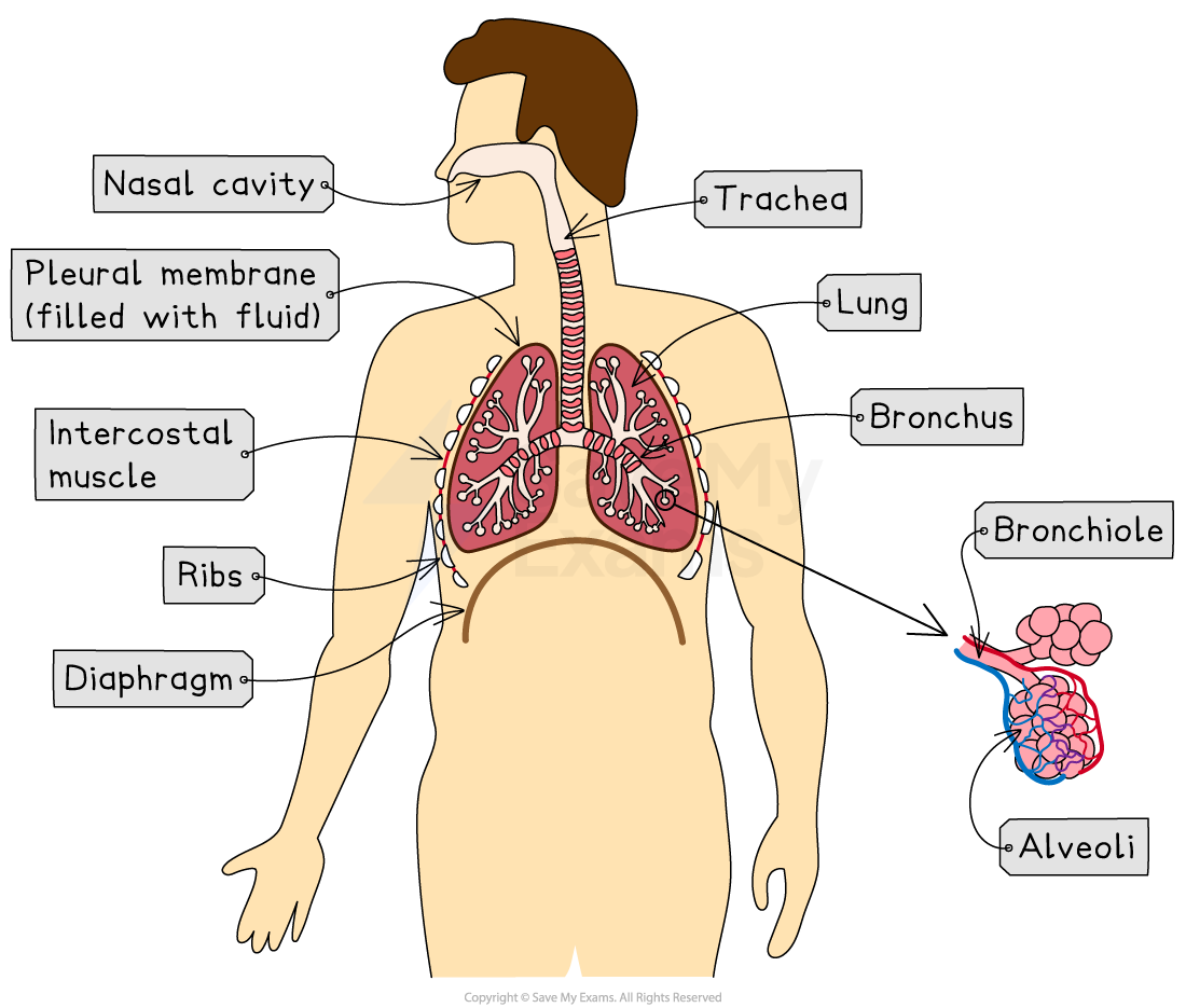
| Structure | What it is / what it does |
|---|---|
| Larynx | The voice box, found at the top of the trachea. Air passes through it when you breathe. |
| Trachea | The windpipe — a single tube that carries air from the throat down into the chest. It is held open by rings of cartilage. |
| Bronchi (singular: bronchus) | The trachea splits into two bronchi — one going into each lung. They carry air from the trachea into each lung. |
| Bronchioles | Each bronchus divides into many smaller tubes called bronchioles, which spread through the lungs like branches of a tree. |
| Alveoli (singular: alveolus) | Tiny air sacs at the ends of the bronchioles. This is where gas exchange takes place between the air and the blood. |
| Associated capillaries | A dense network of very thin blood vessels that surrounds each alveolus. Blood flowing through these capillaries picks up oxygen and releases carbon dioxide. |
5. The Alveoli — The Gas Exchange Surface #
The alveoli are the actual site of gas exchange. Their structure gives them all three features of an efficient gas exchange surface.
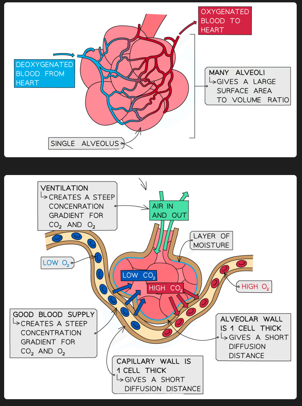
Characteristics of the alveoli #
- Enormous total surface area — there are approximately 700 million alveoli in the two lungs, giving a total surface area of around 70 m² (the size of a tennis court).
- Very thin walls — the alveolus wall is only one cell thick, so gases only need to cross a very short distance.
- Moist inner surface — gases dissolve in this moisture, which allows them to diffuse through the wall.
- Rich blood supply — each alveolus is surrounded by a dense network of capillaries. Blood constantly brings carbon dioxide and takes away oxygen, maintaining a large concentration difference.
How gas exchange happens at the alveoli #
Diffusion moves each gas from where it is at a higher concentration to where it is at a lower concentration:
- Oxygen — higher concentration in the alveolus air than in the blood → diffuses from the alveolus into the blood.
- Carbon dioxide — higher concentration in the blood than in the alveolus air → diffuses from the blood into the alveolus.
6. The Breathing Mechanism #
Structures involved #
You need to identify these on a diagram:
- Ribs — the bony cage that surrounds and protects the lungs.
- External intercostal muscles — muscles between the ribs on the outside. When they contract, the ribs move up and out.
- Internal intercostal muscles — muscles between the ribs on the inside. When they contract, the ribs move down and in.
- Diaphragm — a dome-shaped sheet of muscle below the lungs. When it contracts, it flattens downward.
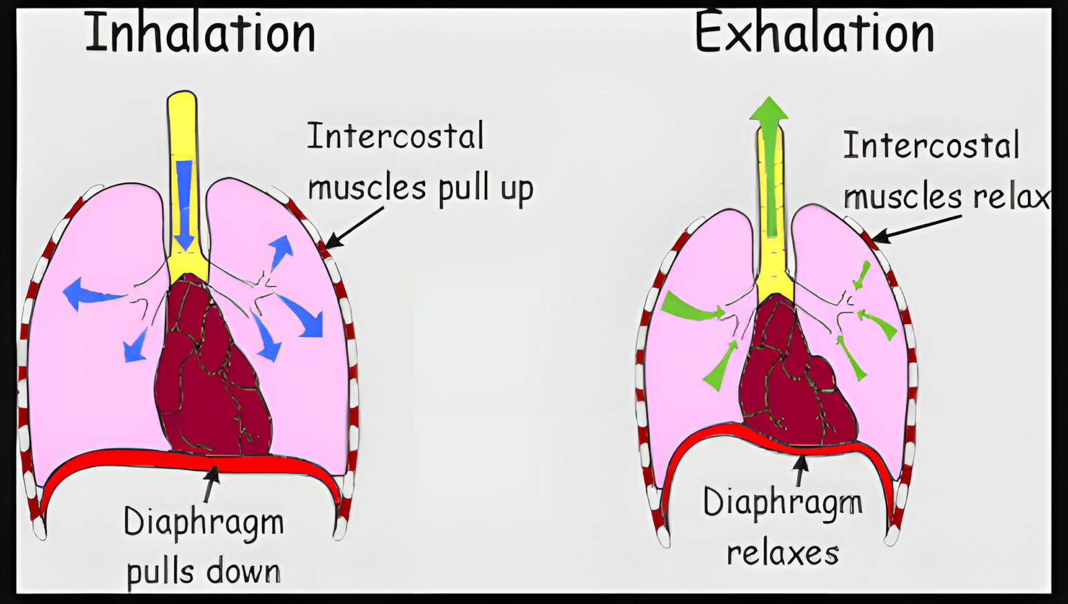
How breathing works — pressure and volume #
The lungs cannot move on their own. They expand or compress because the volume of the chest (thorax) changes, which changes the air pressure inside.
| Inhalation (breathing in) | Exhalation (breathing out) | |
|---|---|---|
| External intercostal muscles | Contract | Relax |
| Internal intercostal muscles | Relax | Contract (in forced exhalation) |
| Ribs | Move up and out | Move down and in |
| Diaphragm | Contracts → flattens | Relaxes → returns to dome shape |
| Volume of thorax | Increases | Decreases |
| Air pressure in lungs | Decreases (below atmospheric) | Increases (above atmospheric) |
| Air movement | Air moves into the lungs | Air moves out of the lungs |
7. Effect of Physical Activity on Breathing #
During exercise, your muscles work harder and need more oxygen. They also produce more carbon dioxide. Your body responds by changing your breathing.
| What changes | During exercise | Why |
|---|---|---|
| Breathing rate (breaths per minute) | Increases | More frequent breaths keep replacing the oxygen used and removing the extra CO₂ produced. |
| Depth of breathing (volume of each breath) | Increases | Each breath brings in and removes a larger volume of air, increasing the total amount of gas exchanged per minute. |
8. Protecting the Gas Exchange System #
The airways are exposed to the air you breathe, which contains dust, bacteria, viruses, and other particles. The lining of the trachea and bronchi has specialised cells that protect the lungs.
| Cell / Substance | What it does | How it protects |
|---|---|---|
| Goblet cells | Produce mucus (a thick, sticky fluid) | Mucus traps dust particles, bacteria, and other pathogens (germs) before they can reach the alveoli. |
| Mucus | A sticky layer that coats the airway lining | Catches pathogens and particles in the air like a sticky trap. |
| Ciliated cells | Cells with tiny hair-like projections called cilia on their surface | The cilia beat in a coordinated wave, sweeping the mucus (with all the trapped particles) up towards the throat, where it is swallowed or expelled. This prevents pathogens from entering the lungs. |
Syllabus Reference — 9.1 Human Gas Exchange #
- Describe the features of gas exchange surfaces in humans, limited to: large surface area, thin surface, good blood and air supply
- State the percentages of the gases in atmospheric air
- Investigate and explain the differences between inspired and expired air
- Identify, on diagrams and images, the larynx, trachea, lungs, bronchi, bronchioles, alveoli and associated capillaries
- State the characteristics of, and describe the role of, the exchange surface of the alveoli in gas exchange
- Identify, on diagrams and images, the ribs, internal and external intercostal muscles and the diaphragm
- Explain the role of the ribs, the internal and external intercostal muscles and the diaphragm in producing volume and pressure changes in the thorax, causing the movement of air into and out of the lungs (breathing)
- Investigate and explain the effect of physical activity on rate and depth of breathing
- Explain the role of goblet cells, ciliated cells and mucus in protecting the gas exchange system from pathogens and particles


