IGCSE Biology | Transport in Humans
Your body is made of billions of cells. Every single one of them needs a constant supply of oxygen, glucose, and other substances — and needs waste products removed. The circulatory system is the transport system that does this job. At the centre of it is the heart, a powerful muscle that pumps blood around your body non-stop, every minute of your life.
1. The Circulatory System #
There are three main parts:
- Heart — the pump that pushes blood through the vessels
- Blood vessels — the tubes that carry blood (arteries, veins, and capillaries)
- Valves — flaps that open and close to stop blood flowing backwards
2. Double Circulation #
Humans have two separate loops of circulation, and blood travels through the heart once in each loop:
Loop 1 — To the Lungs #
Heart → Lungs → Heart
Blood travels from the heart to the lungs to pick up oxygen and drop off carbon dioxide. It then returns to the heart.
Loop 2 — To the Body #
Heart → Body → Heart
Blood travels from the heart to the rest of the body (organs, muscles, tissues) to deliver oxygen. It then returns to the heart.
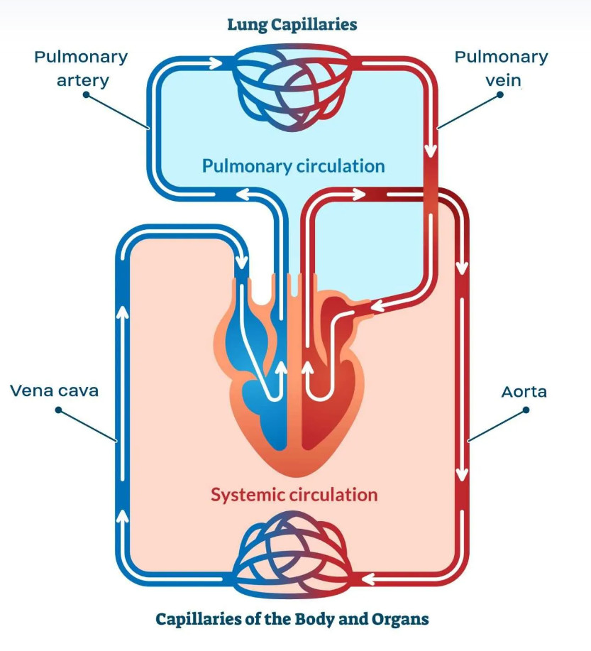
Why is double circulation better? #
Because blood passes through the heart again between the two loops, the heart can pump it at different pressures for each loop:
| Loop | Pressure | Reason |
|---|---|---|
| Pulmonary (to lungs) | Low pressure | The lungs are delicate. High pressure would damage the thin capillary walls where gas exchange happens. |
| Systemic (to body) | High pressure | Blood needs to travel long distances to reach all parts of the body, so it needs a strong push. |
3. Structure of the Heart #
The heart is a muscular organ divided into four chambers. The left and right sides are completely separated by a wall called the septum.
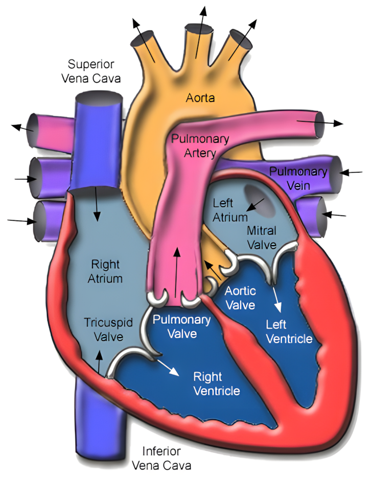
| Structure | What it does |
|---|---|
| Muscular wall | The thick muscle of the heart that contracts to pump blood. |
| Septum | The wall that separates the left and right sides of the heart. This prevents oxygenated and deoxygenated blood from mixing. |
| Right atrium | Upper right chamber. Receives deoxygenated blood returning from the body. |
| Right ventricle | Lower right chamber. Pumps deoxygenated blood to the lungs. |
| Left atrium | Upper left chamber. Receives oxygenated blood returning from the lungs. |
| Left ventricle | Lower left chamber. Pumps oxygenated blood to the whole body. |
| Atrioventricular (AV) valves | Valves between each atrium and ventricle. They stop blood flowing back from the ventricles into the atria. |
| Semilunar valves | Valves at the exits of both ventricles. They stop blood flowing back into the ventricles from the arteries. Found in the pulmonary artery (right) and aorta (left). |
| Coronary arteries | Small arteries that run over the surface of the heart. They supply the heart muscle itself with oxygenated blood and glucose so it can keep contracting. |
4. Thickness of the Heart Walls #
Different parts of the heart have different wall thicknesses, and this matches the amount of force they need to produce.
Left ventricle vs. right ventricle #
Left Ventricle — Thicker wall #
- Pumps blood to the whole body
- Blood must travel a long distance
- Needs high pressure → thicker muscle to contract with more force
Right Ventricle — Thinner wall #
- Pumps blood only to the lungs
- Lungs are nearby and delicate
- Needs lower pressure → thinner muscle wall
Ventricles vs. atria #
Ventricles — Thicker walls #
- Pump blood out of the heart to the lungs or body
- Need strong contractions to push blood a long way
Atria — Thinner walls #
- Only push blood into the ventricles just below them
- Very short distance → much less force needed → thinner walls
5. How the Heart Works #
The heart pumps in a rhythmic cycle. Each heartbeat has two main stages: the atria contracting, then the ventricles contracting.
- Atria fill with blood — Blood flows from the veins into the relaxed atria. The Atrioventricular Valves are open; blood begins to fill the ventricles too.
- Atria contract — Both atria squeeze, pushing the remaining blood through the open Atrioventricular Valves into the ventricles below.
- Ventricles contract — Both ventricles squeeze hard. The pressure forces the Atrioventricular Valves shut (preventing backflow into the atria) and pushes the semilunar valves open. Blood is pushed out into the pulmonary artery (right) and aorta (left).
- Ventricles relax — The pressure in the ventricles drops. The semilunar valves snap shut, stopping blood flowing back from the arteries into the ventricles. The cycle begins again.
The role of valves #
Valves are like one-way doors. They open when pressure pushes them forward and snap shut when pressure tries to push blood backwards. This ensures blood always flows in one direction.
6. Monitoring the Heart #
Doctors use several methods to check how the heart is working:
| Method | What it measures / detects |
|---|---|
| ECG (Electrocardiogram) | Records the electrical signals that trigger each heartbeat. The pattern of peaks and troughs shows whether the heart is beating normally. |
| Pulse rate | Each heartbeat causes a pulse of pressure in the arteries. You can feel this at the wrist or neck. Counting pulses per minute gives the heart rate. |
| Listening to valve sounds | The sounds of a heartbeat are caused by the valves snapping shut. Listening to these sounds shows whether the valves are working correctly. |
7. Physical Activity and Heart Rate #
During exercise, your muscles work harder and need more oxygen and glucose. Your heart responds by beating faster.
Why does heart rate increase during exercise? #
- Contracting muscles use oxygen and glucose more quickly, and produce more carbon dioxide.
- The heart beats faster to speed up blood flow, delivering more oxygen to muscles and removing waste products more quickly.
Method:
- Measure resting heart rate (count pulse for 60 seconds).
- Exercise for a set time (e.g. 2 minutes of stepping or jogging).
- Measure heart rate immediately after stopping.
- Continue measuring every minute until heart rate returns to resting.
- Repeat to get reliable results and calculate an average.
Expected results: Heart rate rises during exercise. After stopping, it gradually returns to the resting rate as demand for oxygen decreases.
8. Coronary Heart Disease (CHD) #
How does it happen? #
Over time, fatty deposits build up inside the walls of the coronary arteries, making them narrower and reducing blood flow.
- The narrowed arteries mean the heart muscle receives less oxygen and glucose.
- If an artery becomes completely blocked, the heart muscle cannot function properly.
Risk factors for CHD #
| Risk Factor | How it increases risk |
|---|---|
| Diet | An unhealthy diet leads to fatty deposits building up in the coronary arteries, increasing the risk of blockage. |
| Sedentary lifestyle | Lack of physical activity leads to weight gain, higher blood pressure, and weaker cardiovascular fitness. |
| Stress | Can raise blood pressure over time, putting extra strain on artery walls and the heart. |
| Smoking | Chemicals in cigarette smoke damage artery walls, making it easier for plaque to form. Smoking also raises blood pressure. |
| Genetic predisposition | Some people inherit a tendency toward high cholesterol or high blood pressure, increasing their risk regardless of lifestyle. |
| Age | Plaque builds up gradually over many years, so risk increases with age. |
| Gender | Males are generally at higher risk of CHD than females. |
9. Reducing the Risk of Coronary Heart Disease #
Two of the most effective ways to reduce the risk of CHD are diet and exercise.
Healthy Diet #
- Eat less fat → reduces fatty deposits building up in the arteries
- Eat more fruit and vegetables
- Maintain a healthy body weight → less strain on the heart
Regular Exercise #
- Strengthens the heart muscle → it pumps more blood with each beat
- Lowers resting blood pressure
- Helps maintain a healthy weight
- Helps reduce the build-up of fatty deposits in the arteries
Syllabus Reference — Section 11: Transport in Humans #
11.1 Circulatory system #
- Describe the circulatory system as a system of blood vessels with a pump and valves to ensure one-way flow of blood
- Describe a double circulation as a system in which blood passes through the heart twice for each complete circuit
- Understand that a double circulation provides a low pressure circulation to the lungs and a high pressure circulation to the body tissues
11.2 Heart #
- Identify the structures of the mammalian heart, limited to: the muscular wall, the septum, the left and right ventricles and atria, atrioventricular and semilunar valves and coronary arteries
- Explain the relative thickness:
(a) of the muscle walls of the left and right ventricles
(b) of the muscle walls of the atria compared to those of the ventricles - Describe the functioning of the heart in terms of the contraction of muscles of the atria and ventricles and the action of the valves in a heartbeat
- State that blood is pumped away from the heart in arteries and returns to the heart in veins
- State that the activity of the heart may be monitored by electrocardiogram (ECG), pulse rate and listening to sounds of valves closing
- Investigate and explain the effect of physical activity on heart rate
- Describe coronary heart disease in terms of the blockage of coronary arteries and state the possible risk factors including diet, sedentary lifestyle, stress, smoking, genetic predisposition, age and gender
- Discuss the role of diet and exercise in reducing the risk of coronary heart disease


